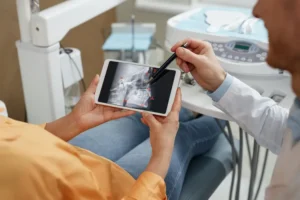
You’ve probably had dental X-rays before. You bite down on that little plastic thing, the machine buzzes, and a few minutes later you’re done. But when it comes to oral surgery and dental implants, those standard X-rays only tell part of the story. That’s where cone beam CT scans come in.
A cone beam CT scan is basically a 3D picture of your mouth, jaws, and sinuses. Unlike regular dental X-rays that flatten everything into a single image, this scan lets us see exactly what’s going on from every angle. We can measure bone height, check nerve locations, spot infections, and plan surgeries with a level of detail that wasn’t possible just a few years ago.
Why Regular X-Rays Aren’t Enough
Standard X-rays are great for finding cavities and checking basic bone levels. But they’re two-dimensional. That means they squish a three-dimensional object into a flat image. You lose depth. You lose perspective. And when you’re planning something like a dental implant, those missing details matter.
Let’s say you need an implant in your lower jaw. There’s a nerve that runs right through that area called the inferior alveolar nerve. If an implant goes too deep and hits that nerve, you could end up with permanent numbness in your lip or chin. With a standard X-ray, we can see the nerve’s general location, but we’re kind of guessing at the exact position. With a cone beam CT scan, we know precisely where that nerve sits within a fraction of a millimeter.
What Happens During the Scan
The good news is that a cone beam CT scan could not be easier. You sit in a comfortable chair while a machine rotates around your head for about 20 to 40 seconds. That’s it. No biting on anything. No weird tastes. No claustrophobic tubes.
The amount of radiation is surprisingly low. In fact, a cone beam scan of your entire mouth uses about the same radiation as a full set of traditional dental X-rays. And it’s far less than a medical CT scan you’d get at a hospital.
How We Use the Information
At East Tennessee Periodontics, we use cone beam CT scans for almost every implant we place. Before Dr. Robert Cain ever picks up a surgical instrument, he’s already mapped out the entire procedure on a computer. He knows exactly where the implant will go, how deep it will be, and whether there’s enough bone to support it.
We also use the scans for sinus lifts, bone grafting, and wisdom tooth extractions. For sinus lifts especially, the scan shows us exactly where the sinus floor sits and how much bone we have to work with. No surprises when we get in there.
The scan also helps us spot problems you might not even know you had. Cysts, tumors, hidden infections, and impacted teeth all show up clearly in 3D. Finding these issues before surgery means we can adjust our plan or refer you to the right specialist before any problems come up.
Better Outcomes for You
Here’s what all this technology really means for you: safer surgeries, fewer surprises, and better results. When we know exactly what we’re walking into, we can avoid nerves, sinuses, and other important structures. Implants placed with cone beam guidance have higher success rates and lower complication rates.
You also spend less time in the chair. Because we’ve already planned everything on the computer, the actual surgery moves faster. Less guesswork means less time with your mouth open.
We Have This Technology in Knoxville
Not every dental office has a cone beam CT scanner. At East Tennessee Periodontics, we invested in this technology because it simply leads to better care for our patients. No referrals to outside imaging centers. No waiting days for results. We scan, we plan, and we treat, all under one roof.
If you’re considering dental implants or any oral surgery procedure, ask about cone beam CT imaging. Or better yet, just give us a call. We’d love to show you how 3D technology can make your treatment safer, smoother, and more predictable.